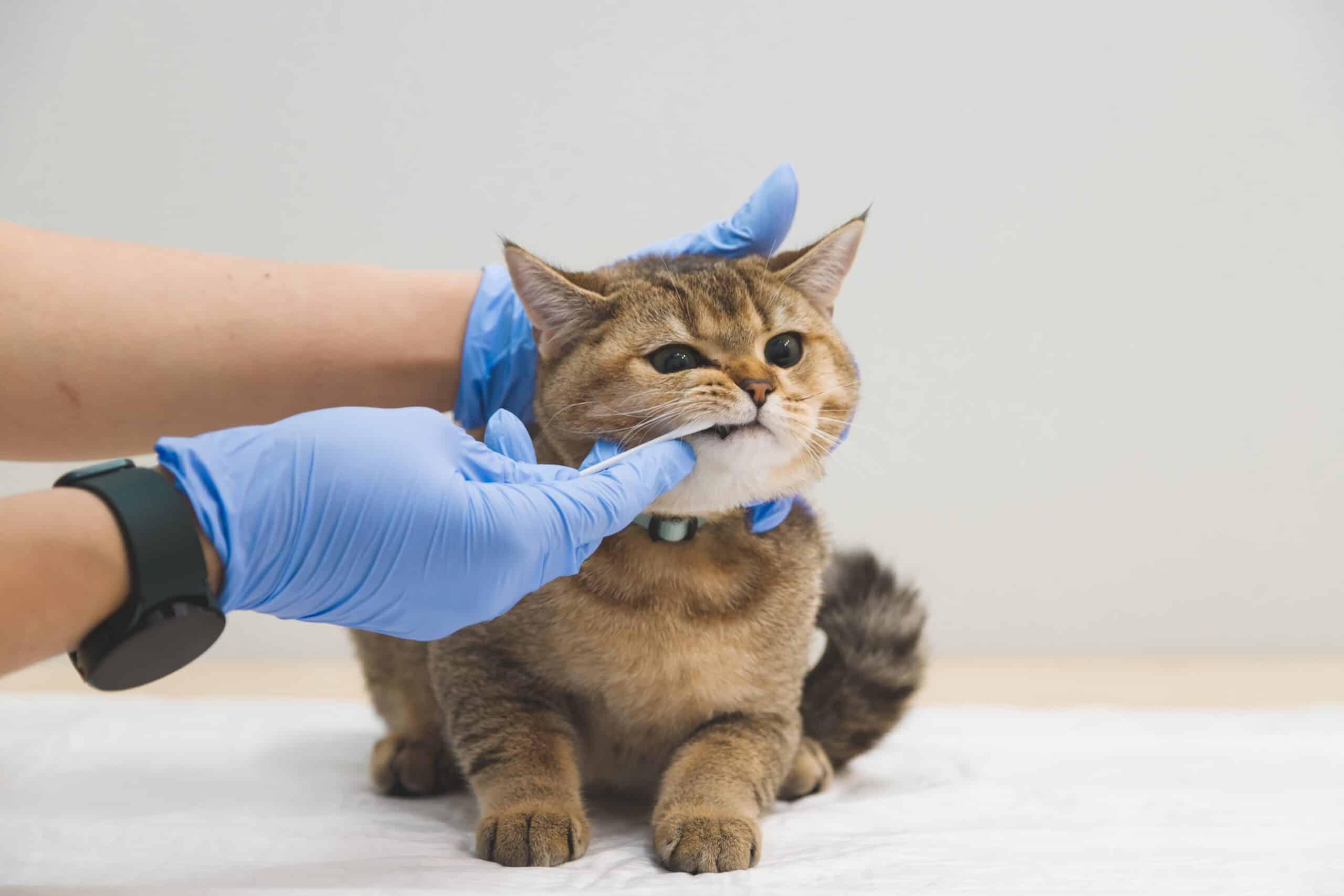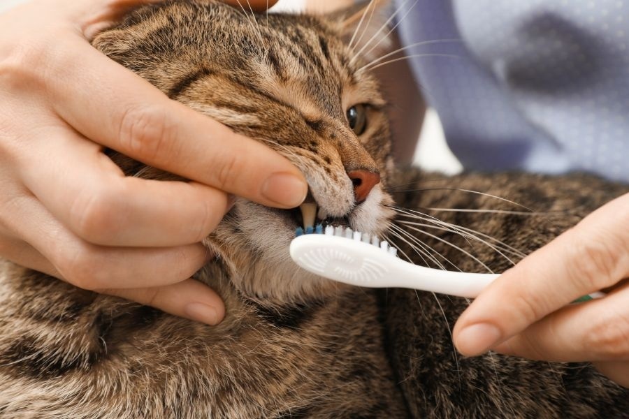
A pet may have clean-looking teeth and still experience significant dental problems beneath the gumline. While visible tartar, bad breath, and red gums can alert pet owners to oral health concerns, some of the most painful conditions remain hidden from view. This is why a professional dental examination involves more than simply looking at the surfaces of the teeth.
Digital dental X-rays give veterinarians a closer look at the structures supporting each tooth. They can reveal problems affecting the roots, surrounding bone, and tissues that cannot be properly evaluated during a quick awake mouth check. For pet parents in Rock Hill, understanding this technology can make it easier to recognize the importance of comprehensive pet dental care. River’s Edge Animal Hospital can help you understand how professional pet dentistry supports your companion’s comfort and oral health.
Why a Visual Dental Examination Is Not Enough
Looking inside a pet’s mouth is an important part of a dental assessment, but it provides only limited information. The visible crown is just one part of a tooth. Beneath the gumline are the roots and supporting structures that help keep the tooth stable.
A substantial portion of a tooth’s structure is hidden below the gums. In some teeth, more than 60 percent may be below the gumline, making a visual examination insufficient for identifying every problem.
For example, a tooth may appear normal from the outside while its root is damaged or the surrounding bone is deteriorating. A pet may also have painful changes beneath the gumline without showing obvious signs during an examination.
Digital intraoral X-rays help reveal these hidden concerns. They provide images of individual teeth and their supporting structures, allowing the veterinary team to evaluate areas that cannot be seen directly.
What Are Digital Dental X-Rays for Pets?
Digital dental radiography is an imaging technique used to examine teeth, roots, and the surrounding jawbone. A small sensor captures images that can be viewed on a computer shortly after they are taken.
Unlike ordinary photographs of the mouth, dental X-rays show structures beneath the surface. They help veterinarians assess the condition of tooth roots, identify changes in bone density, and investigate possible disease affecting the tissues around a tooth.
These images are particularly useful during professional dental cleanings because they help provide a more complete picture of oral health. The veterinarian can compare visible findings with what appears on the X-rays before deciding whether additional treatment is necessary.
Digital imaging can also make it easier to document dental conditions and monitor changes during future examinations.
Hidden Dental Problems That X-Rays Can Reveal
Several oral conditions may develop beneath the gumline without creating obvious changes on the visible surface of a tooth. Digital dental X-rays help identify these problems so they can be evaluated and treated appropriately.
Tooth root abscesses
A tooth root abscess occurs when infection develops around the root of a tooth. It may be associated with a damaged tooth, advanced dental disease, or infection within the tooth.
A pet may experience pain while chewing, swelling around the face, or changes in eating habits. However, symptoms are not always obvious. X-rays can reveal changes around the root that suggest infection or other underlying damage.
Bone loss from periodontal disease
Periodontal disease affects the tissues that support the teeth. As the condition progresses, inflammation and infection can damage the structures around the roots, including the supporting bone.
Digital images help veterinarians assess bone levels and identify areas of deterioration. This information can help determine how severely a tooth is affected and whether it can be maintained or requires treatment.
Tooth resorption in cats
Tooth resorption is a painful condition in which a tooth gradually breaks down. It is particularly common in cats and may affect different parts of the tooth.
Some affected teeth may look relatively normal during a visual examination, even though significant damage is occurring beneath the gums. Dental X-rays help identify the extent and pattern of resorption, which guides treatment decisions.
Retained roots and hidden fractures
Occasionally, a tooth may break or sustain damage below the visible crown. In other cases, a previous extraction may leave root fragments behind. These issues may not be apparent during a routine visual check.
Dental imaging helps the veterinarian investigate the full tooth structure and determine whether additional treatment is needed.
Why Dental X-Rays Are Usually Taken Under Anesthesia
Taking detailed dental X-rays requires accurate positioning of the sensor and the pet’s head. The animal must remain still while images are captured from different angles.
Anesthesia allows the veterinary team to perform a thorough oral examination and obtain diagnostic images without causing unnecessary stress or discomfort. It also makes it possible to clean beneath the gumline, assess individual teeth, and address dental problems during the same procedure when appropriate.
During anesthetic dental care, the veterinary team follows a planned protocol that may include:
- Reviewing the pet’s medical history and performing a pre-procedure examination.
- Recommending blood screening when appropriate.
- Selecting an anesthetic plan based on the pet’s health.
- Protecting the airway with an appropriate breathing tube.
- Monitoring vital signs throughout the procedure.
- Using dental instruments and imaging equipment to evaluate oral health.
Anesthesia carries risks, but pre-procedure assessment and continuous monitoring help the veterinary team manage those risks. Your veterinarian can explain the benefits and considerations based on your pet’s age and medical condition.
Why Anesthesia-Free Dental Cleaning Has Important Limitations
Anesthesia-free dental cleaning may appear convenient because the pet remains awake. However, it generally focuses on removing visible tartar from the exposed surfaces of the teeth.
It does not allow the veterinary team to perform a complete examination beneath the gumline, obtain properly positioned intraoral X-rays, or thoroughly clean the areas where periodontal disease develops.
A pet may also move, resist handling, or experience discomfort when someone works inside the mouth. This makes it difficult to examine every tooth and safely address painful areas.
Removing visible tartar does not necessarily resolve infection, root damage, bone loss, or tooth resorption. A surface-only cleaning can therefore leave significant dental disease undetected.
Professional pet dentistry under anesthesia provides the conditions needed for a more complete assessment and treatment. It focuses on the health of the entire tooth, not just the part that can be seen.
How Digital Imaging Guides Dental Treatment
Dental X-rays do more than identify problems. They also help veterinarians decide which treatment may be appropriate.
If imaging reveals advanced bone loss, a root abscess, or severe tooth resorption, the veterinarian may recommend extraction. If a tooth has a condition that can be treated or monitored, the veterinary team can explain the available options.
Imaging is also useful after certain dental procedures. Post-treatment images may help confirm that affected root structures have been addressed appropriately.
The treatment plan depends on the findings, the pet’s overall health, and the condition of each tooth. Your veterinarian should explain the results and discuss recommended next steps before treatment whenever possible.
Signs Your Pet May Need a Dental Evaluation
Pets do not always show obvious signs of oral pain. Changes in routine behavior may be the first indication that something is wrong.
Watch for:
- Persistent bad breath.
- Red, swollen, or bleeding gums.
- Dropping food while eating.
- Chewing on one side of the mouth.
- Pawing at the face or mouth.
- Increased drooling.
- Reluctance to eat hard food or chew toys.
- Changes in behavior when the face is touched.
Even if your pet continues eating normally, dental disease may still be present. Regular veterinary examinations help identify concerns before they progress.
Give Your Pet a More Complete Dental Examination in Rock Hill
Healthy teeth involve much more than a clean-looking smile. Problems beneath the gumline can affect roots, supporting bone, and surrounding tissues without being visible during an ordinary mouth check. Digital dental X-rays help veterinarians identify these concerns and make informed treatment recommendations.
If your dog or cat is due for a dental evaluation, contact River’s Edge Animal Hospital in Rock Hill. Schedule an appointment to discuss your pet’s oral health, professional cleaning options, and whether digital dental imaging may be appropriate. A thorough approach to pet dental care can help identify hidden problems and support long-term comfort.
Frequently Asked Questions
Q1. Why do pets need dental X-rays if their teeth look clean?
Ans: Dental disease can develop beneath the gumline, affecting roots and supporting bone without creating obvious surface changes. X-rays help veterinarians identify these hidden problems.
Q2. Can digital dental X-rays detect tooth root infections in pets?
Ans: Yes. Dental images can reveal changes around tooth roots that may indicate an abscess or other underlying damage. The veterinarian uses these findings alongside the oral examination to determine appropriate treatment.
Q3. Are dental X-rays safe for dogs and cats?
Ans: Dental X-rays use controlled amounts of radiation to produce diagnostic images. The veterinary team follows appropriate imaging protocols and considers the pet’s individual needs when recommending the procedure.
Q4. Why is anesthesia needed for professional pet dental imaging?
Ans: Anesthesia helps pets remain still while the veterinary team positions the imaging sensor and examines the entire mouth. It also allows thorough cleaning beneath the gumline and treatment of dental problems when needed.
Q5. How often should my pet have dental X-rays?
Ans: The need for dental imaging depends on your pet’s age, oral health, previous dental findings, and clinical examination. Your veterinarian can recommend an appropriate schedule based on individual needs.